Rare skeletal muscle metastasis from sclerosing sub-type of papillary thyroid cancer (#413)
Thyroid cancer is the most common endocrine malignancy with 820,000 cases annually [1]. Distant metastases most commonly affect the lungs, bone or brain, with other organs involved in <5% of cases [2]. Skeletal muscle metastases are exceedingly rare, with a literature review from 1907-2017 identifying only 58 cases, with these patients having a poorer prognosis compared to other sites of distant metastases [3].
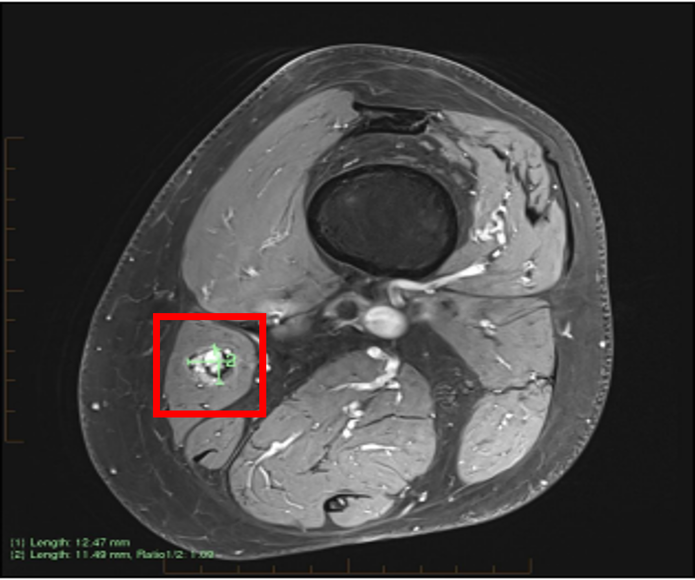
A 62-year-old man presented for ongoing surveillance following high-risk sclerosing subtype of papillary thyroid cancer. He was initially treated with total thyroidectomy, bilateral neck dissection and 6GBq radioactive iodine (RAI) in 2018. Subsequent imaging confirmed radioactive iodine refractory disease. He underwent three further neck dissections for loco-regional disease. He developed asymptomatic pulmonary metastases in 2023. Despite ongoing TSH suppression, he was found to have a rising thyroglobulin from 2.7ug/L to 14.6ug/L over 6 months in 2025. FDG-PET/CT scan demonstrated stable disease in the lungs and neck, but a new glucose-avid lesion in his left adductor muscle (SUV = 6.3) (Figure 1). MRI thigh showed a suspicious 13x12x11mm lesion in the distal sartorius musculature with intralesional haemorrhage (Figure 2). Ultrasound guided biopsy of the lesion was performed, with immunohistochemistry (positive pan CK TTF-1, thyroglobulin and BRAF stain) and positive thyroglobulin washout confirming metastatic thyroid carcinoma. The patient remains reluctant to commence systemic therapy given he is asymptomatic with an excellent ECOG status. Next-generation sequencing is pending to determine suitability for targeted therapy given ongoing progression and the association with poorer prognosis in the literature.
This case illustrates the potential for thyroid cancer to metastasise to rarer sites, and the importance of histopathological confirmation. Patients with skeletal metastases may warrant more aggressive management given the association with reduced survival compared to other sites of distant metastases.
Figure 1: FDG-PET left adductor lesion
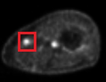
Figure 2: MRI left sartorius lesion
- Chen DW, Haymart MR. Unravelling the rise in thyroid cancer incidence and addressing overdiagnosis. Nat Rev Endocrinol. 2026 Jan;22(1):10-20. doi: 10.1038/s41574-025-01168-y. Epub 2025 Sep 3. PMID: 40903496; PMCID: PMC12814954.
- Talebpour M, Zabihi Mahmoudabadi H, Najjari K, Hajebi R, Rahimpour E, Bahreini M. Distant Metastasis as the First Presentation of Thyroid Carcinoma. Indian J Otolaryngol Head Neck Surg. 2023 Jun;75(2):1020-1023. doi: 10.1007/s12070-022-03343-3. Epub 2022 Dec 8. PMID: 37275079; PMCID: PMC10235276.
- Herbowski L. Skeletal muscle metastases from papillary and follicular thyroid carcinomas: An extensive review of the literature. Oncol Lett. 2018 May;15(5):7083-7089. doi: 10.3892/ol.2018.8216. Epub 2018 Mar 8. PMID: 29731874; PMCID: PMC5920897.