A Case of ACTH dependent paraneoplastic Cushing’s syndrome in metastatic small cell cervical cancer (#417)
Ectopic paraneoplastic Cushing's syndrome is uncommon and even less so in small cell cervical cancer (SCCC). We report a case of ACTH-dependent paraneoplastic Cushing’s syndrome in a 46-year-old female with metastatic SCCC as well as adjuvant treatment with osilodrostat for management of progressively aggressive clinical features.
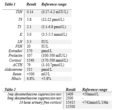
A 46-year-old female of Afghani background presented with lethargy, weight loss and a four-month history of abdominal distension and heavy menstrual bleeding. Medical background was significant for obstructive sleep apnoea, treated hypertension and schizoaffective disorder. Initial investigations showed metabolic alkalosis and hypokalaemia (pH 7.48, carbon dioxide 41, potassium 3.0mmol/L). A large uterine density (24x17cm) with paraaortic lymphadenopathy was identified in commuted tomography. Uterine mass biopsy diagnosed small cell cervical cancer (Cam5.2 and synaptophysin positive, CD56 negative, Ki-67 80%, ACTH negative, CRH not available). Carboplatin/etoposide chemotherapy was commenced. A finding of central hypothyroidism prompted further pituitary workup with a finding of grossly elevated ACTH levels (Table 1).
She initially responded to chemotherapy with downtrending ACTH levels (46pmol/L) and normalization of thyroid function (TSH 3mUI/L, T4 14mIU/L). However, recurrent admissions with hypokalaemia delayed further chemotherapy, and her malignancy progressed aggressively. Cortisol failed to suppress with 1 mg and 8 mg dexamethasone suppression testing (Table 2). Twenty-four-hour urinary free cortisol collection was delayed but was subsequently profoundly elevated (Table 2). Clinically, she developed worsening glycaemic control, weight gain, and classical Cushingoid features including a rounded plethoric face, dorsocervical fat pad, and proximal myopathy. Osilodrostat 1 mg twice daily was commenced and uptitrated to 5 mg twice daily, improving urinary free cortisol levels. Unfortunately, repeat computed tomography imaging demonstrated further aggressive disease progression, and chemotherapy was ceased because of this and her poor ECOG status. She passed away four weeks later. We posit that earlier treatment with osilodrostat may have mitigated chemotherapy delays and improved survival outcomes.